
Release date: 2022.10.31
In connection with Pink Ribbon Month in October, which calls for early breast cancer screening, we will summarize breast cancer screenings in three parts. In Part 3, we will introduce CT (computed tomography), MRI (magnetic resonance imaging), bone scintigraphy, and PET (positron emission tomography) examinations that examine the spread and metastasis of breast cancer.
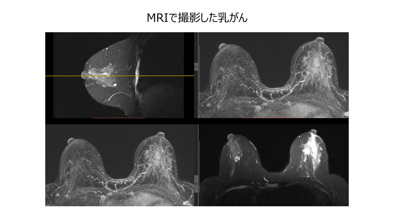
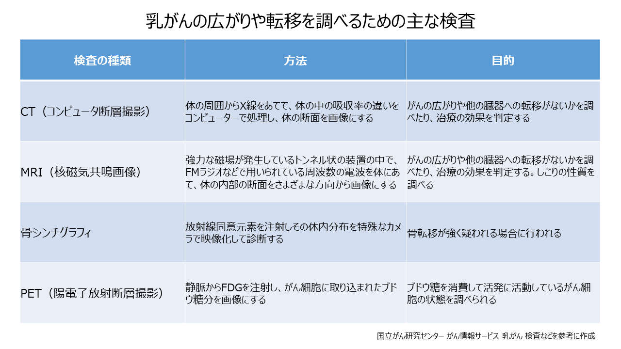
A CT scan is a type of X-ray imaging that uses sliced images of the body to make a diagnosis. It is possible to check the spread of cancer in the breast and the presence or absence of distant metastases that spread to other organs. Breast cancer can metastasize to distant lymph nodes in the neck, bones, lungs, liver, and brain. Magnetic resonance imaging (MRI) makes a diagnosis by imaging signals emitted from the body in response to magnetism. It is possible to observe every tomographic plane of the body. It uses magnets and radio waves instead of X-rays, so you don't have to worry about radiation exposure. Similar to CT, it is possible to examine the spread of cancer within the breast and the properties of lumps. Painless MRI breast cancer screening (Duibus Search), which is considered a type of MRI examination, is suitable for those who shy away from screening mammograms because of the pain involved. The examination is performed by lying face down on a bed that has been hollowed out in the shape of a breast, so there is no pain because the chest is not compressed. This test does not affect the amount of breast tissue, so the cancer detection rate does not decrease even in dense breasts, and radiation exposure is zero.
Bone scintigraphy is a test that images gamma rays emitted from bone tissue in the body by intravenously injecting a drug containing a radioactive isotope that has the property of being concentrated in bones throughout the body. This is done to check if the breast cancer has spread to the bone. After the injection, images will be taken after the drug has fully accumulated in the bone, but it will take at least 2 hours to accumulate.
PET scans are also used to check for metastasis to other organs. The test method involves injecting a testing agent called FDG, which is very similar to glucose, into the body and imaging the distribution of glucose taken up by cancer cells. Many breast cancers take up FDG well, and it is said that the diagnostic ability is high in organs such as bone, lung, and liver, where the frequency of breast cancer metastasis is high. It may be used when other tests, such as CT or MRI, do not provide a clear diagnosis.
CT examination, MRI examination, bone scintigraphy, and PET examination are performed not only before deciding the treatment policy, but also to confirm the effect of drug therapy and to check for recurrence.
Tumor markers are a method of testing the amount of proteins produced and secreted by cancer cells using blood and urine, and are used as clues for cancer diagnosis. There are no tumor markers for staging. However, tumor markers are routinely used as a means of understanding the condition of cancer recurrence and confirming the efficacy of treatment. CEA, CA15-3, and NCC-ST-439 are widely used as tumor markers for breast cancer. If the tumor marker is abnormally high, it is judged as positive, and if it is low, it is judged as negative.


 URL copied
![]()
MEDIUS Group is developing a business centered on the sale of medical equipment. We (Medical + us) involved in medical care also want to play the role of an information source (Media) that delivers useful information for the medical field and people's healthy tomorrow.


The challenge of home medical care that "supports daily life" - A "triple win" for patients, medical institutions, and the government will ensure the future of an aging society...

Supporting the career advancement of female doctors Introduce career counseling to increase the number of female doctors aiming to become specialists...

The new value of medical care is to improve the happiness of patients and medical professionals by providing medical services that emphasize "emotional value"

Genomic medicine: the present and future expectations

Supporting regional medical care in disaster-prone Japan: Japan Medical Association Disaster Medical Team (JMAT)