ASOURCE®NAVI

公開日:2024.04.12
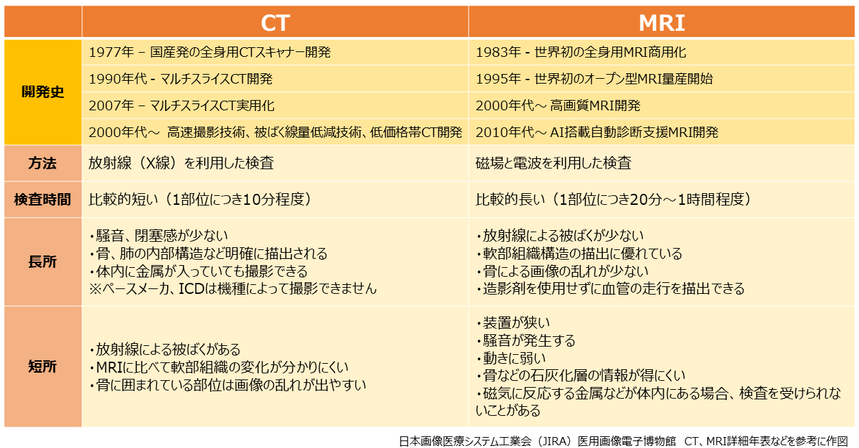
画像診断は、医療において必要不可欠なものとなっていますが、日本は、X線を利用して体内の状態を断面像として描写するCT(Computed Tomography コンピュータ断層撮影)や強い磁石と電波を利用して体内の状態を断面像として描写するMRI(Magnetic Resonance Imaging 磁気共鳴画像)の研究開発において、世界の先導的な役割を果たしてきました。こうした高度な画像診断技術は、疾患の正確な診断と早期発見に大きく貢献しています。
CTは、被ばく線量が低く、撮影時間が短いことが特徴で、骨や石灰化など、硬い組織の画像化に優れています。そのため、外傷や腫瘍の診断、血管造影検査、仮想内視鏡検査など、幅広い分野で活用されています。
CTは1970年代前半に実用化されて以来、約40年にわたる技術革新によって飛躍的に進化を遂げてきました。1990年頃に患者を移動させながら連続撮影するヘリカルスキャンが実用化され、その後、患者を移動させることなく1回転で1つの臓器をスキャンし、連続回転スキャンにより臓器の動きを捉えるマルチスライスCTが開発されました。マルチスライスCTは256列(列:体軸方向の検出器の数を指し、一回転で取得できる画像の枚数。列数が多いほど一回転で広範囲を撮影できる)のプロトタイプを経て、2007年に320列マルチスライスCTが実用化されました。これにより、従来機に比べて冠動脈の形状や血流の詳細な観察能力が大幅に向上しました。
日本のCT技術は、高速撮影技術、被ばく線量低減技術、低価格帯CTなどにおいて世界的に高い評価を受けています。高速撮影技術では、短時間に心臓などの動きを捉え、鮮明な画像を取得できます。被ばく線量低減技術では、従来に比べて患者の被ばく線量を大幅に低減することが可能です。また、低価格帯CTも開発されており、発展途上国での普及に貢献しています。
さまざまな方向からの断面画像を取得できることが、MRI検査の大きな特徴です。放射線を使わない、造影剤を使わなくても血管を撮影でき、骨による影響を受けにくいという特長があり、脳腫瘍や脳梗塞、がんなどを発見するための検査や、子宮や卵巣の状態を調べる検査等に活用されています。
人体内の水素原子核の磁気共鳴を利用して画像を生成するMRIの研究開発は、1970年代から始まり、1980年代に実用化されています。MRIは高磁場であるほど高画質となり、磁場の強さ(磁束密度)はT(テスラ)という単位で表されます。1983年に開発された全身用MRIは0.15Tで、現在の臨床で主流となっている1.5Tあるいは3Tよりも一桁小さいものでしたが、基本的な構成は現在の最新MRIとほぼ同じでした。その後、1995年に世界初のオープン型MRIの量産が開始されました。オープン型MRIは、広い開口部を持ち、患者がトンネル内に閉じ込められることなく検査を受けることができます。これにより、閉所恐怖症や不安を抱える患者にとって、より快適な検査環境を提供することができます。
日本のMRI技術は、世界で初めて商用化された全身用MRIやオープン型MRI、高画質MRI、AI搭載自動診断支援MRIなど、革新的な技術で知られています。1983年に開発された全身用MRIは、現在のMRIの基礎を築きました。また、1995年に量産開始されたオープン型MRIは、閉所恐怖症患者にも快適な検査環境を提供しました。グラジエントコイル技術を用いた高画質MRIは、従来の10倍以上の高精度を実現しています。さらに心電図同期撮影や並列イメージングなどの高速撮影技術も発達しました。そして、AI搭載自動診断支援MRIは、診断精度向上と医師の負担軽減に貢献しています。
CTとMRIの研究開発は、今後も被ばく線量のさらなる低減、高速撮影技術の発展、AIによる画像診断支援、小型・軽量化など、様々な分野で進められていくと考えられます。また、MRIにおいては新しい計測手法の開発など、未知の機能の探索も期待されています。日本のCT・MRI研究開発は、これからも医療の発展に貢献することが期待されます。


 URL copied
![]()
メディアスグループは、医療機器の販売を中心とした事業を展開しています。医療に携わる私たち(Medical+us)は、医療現場や人々の健康的な明日へ役立つ情報をお届けする情報発信源(Media)の役割も果たしていきたいと考えています。